The 9th Edition of Radiation Protection in Medical Radiography serves as a cornerstone for understanding radiation safety, simplifying complex concepts for both novice and advanced practitioners alike.
1.1. Overview of the 9th Edition
The 9th Edition of Radiation Protection in Medical Radiography provides a comprehensive update on radiation safety principles, emphasizing practical applications and emerging technologies in medical imaging. It simplifies complex concepts, making it accessible for both students and professionals. Key updates include discussions on dose reduction strategies, advancements in digital radiography, and the integration of artificial intelligence in radiation dose management. The edition also highlights the importance of the ALARA principle and its implementation in modern radiographic practices. With a focus on patient safety and regulatory compliance, this edition serves as an essential resource for understanding radiation protection in the evolving field of medical radiography.
1.2. Importance of Radiation Safety in Medical Imaging
Medical imaging relies heavily on ionizing radiation, which, while essential for diagnosis, poses risks to patients and staff. Radiation safety is critical to minimize exposure while maintaining image quality. The 9th Edition emphasizes balancing diagnostic benefits with potential risks, ensuring patient protection without compromising care; Proper radiation safety protocols reduce long-term health risks, such as cancer, associated with excessive exposure. By adhering to guidelines, healthcare professionals can optimize dose reduction strategies, ensuring safer practices. This edition underscores the importance of education and training in radiation safety, equipping radiologic technologists with the knowledge to protect patients and themselves effectively in the evolving field of medical radiography.
1.3. Key Updates in the 9th Edition
The 9th Edition of Radiation Protection in Medical Radiography introduces significant updates to enhance understanding and application of radiation safety principles. It simplifies complex concepts, making them accessible to both students and experienced professionals. New chapters focus on advanced technologies like digital radiography and cone-beam computed tomography (CBCT), emphasizing dose reduction strategies. Updated guidelines on fetal protection andgonadal shielding are included, alongside practical case studies to illustrate real-world applications. The edition also incorporates the latest regulatory requirements and advancements in radiation measurement devices. Additionally, enhanced training tools, such as test banks and downloadable workbooks, support comprehensive learning. These updates ensure the text remains a vital resource for modern radiologic practice.
Fundamental Principles of Radiation Protection
Medical imaging relies on ionizing radiation, such as X-rays and CT scans, to produce images. Understanding radiation sources and measurement concepts is crucial for safe practices, ensuring patient and staff safety.
2.1. Types of Radiation and Their Interactions
In medical radiography, ionizing radiation, such as X-rays and gamma rays, is commonly used to produce diagnostic images. These types of radiation possess high energy, enabling them to penetrate tissues and interact with matter.
When radiation interacts with materials, it can undergo processes like absorption, scattering, or attenuation. Understanding these interactions is crucial for optimizing image quality and minimizing exposure. Photon radiation, including X-rays, is the primary type used in radiography, while particle radiation (e.g., alpha, beta particles) is less common but still significant in specific applications.
2.2. Sources of Radiation in Medical Radiography
In medical radiography, the primary source of radiation is the X-ray tube, which produces X-rays through the acceleration and deceleration of high-energy electrons. These tubes are designed to generate controlled amounts of radiation for imaging purposes.
Additional sources include radioactive materials used in certain diagnostic or therapeutic procedures, such as technetium-99m in nuclear medicine. Proper shielding and storage of these sources are critical to minimize exposure. The 9th Edition emphasizes the importance of maintaining equipment and following protocols to ensure radiation is generated and contained safely, reducing risks to patients and staff.
2.3. Basic Concepts of Radiation Measurement
Understanding radiation measurement is crucial for ensuring safety in medical radiography. Key concepts include ionizing radiation, which has enough energy to remove electrons from atoms, potentially causing biological damage. The dose refers to the amount of radiation absorbed by a tissue, measured in grays (Gy) or sieverts (Sv). Exposure measures radiation in the air, while absorbed dose reflects energy deposited per unit mass. The dose equivalent accounts for radiation type and biological effect, using weighting factors. These metrics guide safe practices, helping minimize risks while optimizing image quality. Accurate measurement ensures compliance with safety standards, protecting both patients and staff.

Patient Radiation Protection
Patient radiation protection focuses on minimizing exposure while ensuring diagnostic image quality. Techniques include proper shielding, collimation, and using the lowest necessary dose for accurate imaging.
3.1. Principles of Patient Shielding
Patient shielding is a critical component of radiation protection, aiming to minimize exposure to ionizing radiation while maintaining diagnostic image quality. Shielding involves using materials like lead aprons, thyroid collars, and gonadal shields to block scattered radiation. Proper placement and selection of shielding materials are essential to ensure effectiveness without compromising the radiographic image. The 9th edition emphasizes the importance of tailored shielding strategies based on patient anatomy and procedure type. It also highlights the need for regular inspection of shielding equipment to ensure integrity and compliance with safety standards. By prioritizing shielding, healthcare providers can significantly reduce radiation doses to sensitive tissues, aligning with the ALARA principle.
3.2. Minimizing Patient Dose with Proper Techniques
Minimizing patient radiation dose is achieved through precise radiographic techniques that balance image quality with radiation safety. Proper use of exposure factors, such as kilovoltage (kVp) and milliamperage-seconds (mAs), ensures optimal image quality while reducing dose. Collimation, or restricting the X-ray beam to the desired anatomical area, minimizes scattered radiation and reduces patient exposure. Accurate patient positioning and the use of high-speed imaging receptors further enhance efficiency. Avoiding repetitive exposures and ensuring proper equipment maintenance are also critical. These strategies align with the ALARA principle, emphasizing the lowest reasonable radiation levels while maintaining diagnostic accuracy, as outlined in the 9th edition guidelines.
- Use appropriate exposure factors to optimize image quality and dose.
- Collimate the X-ray beam to the area of interest.
- Ensure accurate patient positioning to reduce retakes.
- Utilize high-speed receptors for dose efficiency.
3.3. Thyroid and Gonadal Protection
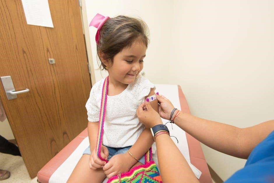
Protecting the thyroid gland and gonads from radiation exposure is critical due to their high sensitivity. The thyroid gland is particularly vulnerable to radioactive iodine, necessitating the use of thyroid collars during procedures. Gonadal protection involves shielding the reproductive organs with lead aprons or gonadal shields to minimize radiation exposure, especially in younger patients. These measures are essential for reducing the risk of genetic mutations and thyroid disorders. The 9th edition emphasizes the importance of assessing the necessity of gonadal shielding on a case-by-case basis to avoid obstructing the diagnostic field. Adherence to these guidelines ensures compliance with radiation safety standards and the ALARA principle.
- Use thyroid collars to protect the thyroid gland.
- Shield gonads with lead aprons or gonadal shields when appropriate.
- Assess the need for shielding to avoid diagnostic obstruction.
- Adhere to guidelines to minimize radiation risk to sensitive tissues.

Radiation Biology and Risk Assessment
Radiation biology examines how ionizing radiation interacts with living cells, causing DNA damage and potential health risks; This section explores radiation-induced biological effects and risk assessment methods in medical imaging.
4.1. Biological Effects of Ionizing Radiation
Ionizing radiation interacts with living tissues, causing biological effects by damaging DNA and other cellular components. This damage can lead to chromosomal abnormalities, mutations, and disruptions in cellular function. Acute exposure may result in deterministic effects, such as radiation burns or organ failure, while chronic low-dose exposure increases the risk of stochastic effects, like cancer or genetic disorders. The severity of these effects depends on the radiation dose, duration of exposure, and sensitivity of the exposed tissue. Understanding these biological interactions is critical for developing strategies to minimize risks and protect patients and professionals in medical radiography. This knowledge underpins effective radiation safety practices.
4.2. Assessing Radiation Risks in Medical Imaging
Assessing radiation risks in medical imaging involves evaluating the potential harm caused by ionizing radiation exposure. This includes calculating effective doses, comparing them to established dose limits, and weighing the benefits of diagnostic imaging against possible risks. Tools like dose-length products and effective dose estimates help quantify exposure. Patient-specific factors, such as age and medical history, are considered to personalize risk assessments. Guidelines from organizations like the National Council on Radiation Protection and Measurements (NCRP) and the International Commission on Radiological Protection (ICRP) provide frameworks for these evaluations. Accurate risk assessment ensures that imaging procedures are optimized, minimizing radiation exposure while maintaining diagnostic quality. This balance is essential for patient safety and ethical practice in medical radiography.
4.3. Dose-Response Relationships
Dose-response relationships describe how biological effects vary with radiation exposure levels. In radiation protection, understanding these relationships is critical for setting safety standards. Deterministic effects, such as radiation burns, exhibit a threshold dose below which they do not occur. Stochastic effects, like cancer induction, show no threshold and increase in probability with higher doses. These relationships help quantify risks and establish exposure limits. They also guide the optimization of imaging protocols to balance diagnostic benefits with radiation risks. Accurate dose-response models enable better risk-benefit analyses, ensuring patient safety while maintaining image quality. This knowledge is vital for advancing radiation protection practices in medical radiography.

Radiation Detection and Measurement
Accurate radiation detection and measurement ensure safety, employing devices like dosimeters and Geiger counters to assess exposure levels, crucial for regulatory compliance and patient protection.
5.1. Devices for Measuring Radiation Exposure
In medical radiography, various devices measure radiation exposure to ensure safety. Personal dosimeters, like thermoluminescent dosimeters (TLDs) and digital dosimeters, monitor individual exposure. Geiger counters detect radiation levels in real-time, while ion chambers measure exposure rates. Film badges provide a permanent record of cumulative exposure. Scintillation detectors offer precise measurements in high-dose environments. These devices help maintain regulatory compliance, ensure patient safety, and verify equipment performance. Proper calibration and use are essential for accurate data. The 9th Edition emphasizes selecting the right device for specific procedures to optimize radiation monitoring and minimize risks effectively.
5.2. Dosimetry in Medical Radiography
Dosimetry in medical radiography involves the measurement and calculation of radiation doses received by patients and staff. It ensures radiation levels are optimized for diagnostic quality while minimizing risks. Personal dosimeters, like thermoluminescent dosimeters (TLDs) and digital dosimeters, are used to monitor individual exposure. Environmental dosimetry measures radiation levels in specific areas. Accurate dosimetry is critical for adhering to the ALARA principle, ensuring doses are as low as reasonably achievable. Modern dosimetry systems provide detailed data, enabling precise risk assessments and compliance with safety standards. The 9th Edition emphasizes the integration of advanced dosimetry techniques to enhance radiation safety and patient care.
5.3. Quality Control in Radiation Measurement
Quality control in radiation measurement ensures accuracy and consistency in monitoring radiation exposure. It involves regular calibration of dosimetry devices, such as ion chambers and thermoluminescent dosimeters, to maintain reliability. Proper maintenance and testing of equipment prevent errors in radiation detection. Standardized protocols are implemented to ensure consistent measurements across different devices and facilities. Routine checks and self-testing of dosimeters are essential to verify their functionality. Documentation of calibration and testing results is critical for compliance with safety standards. Any deviations or malfunctions must be addressed promptly to maintain the integrity of radiation measurements. Adherence to these quality control measures ensures accurate radiation monitoring, safeguarding patients and personnel while optimizing diagnostic outcomes.

Radiation Protection in Specific Medical Procedures
Addresses radiation safety measures tailored to specific medical imaging procedures, ensuring optimal patient protection while maintaining diagnostic image quality through procedure-specific protocols and safeguards.
6.1. Radiation Safety in Dental Radiography
Radiation safety in dental radiography focuses on minimizing exposure while obtaining diagnostic-quality images. Key protocols include using the lowest appropriate X-ray beam energy, adequate filtration, and proper shielding. Lead aprons and thyroid collars are essential for patient protection. Digital imaging systems are preferred as they reduce radiation doses significantly. Regular maintenance of equipment ensures optimal performance and safety. Training in radiation safety is crucial for dental professionals to adhere to guidelines and minimize unnecessary exposure. Patient-specific techniques, such as avoiding unnecessary retakes, further enhance safety. These measures collectively ensure that radiation risks are minimized while maintaining the diagnostic value of dental radiographs.
6.2. Protecting the Fetus in Radiographic Procedures
Protecting the fetus in radiographic procedures is critical due to the sensitivity of developing tissues to ionizing radiation. The primary goal is to minimize fetal exposure while ensuring diagnostic image quality. Key measures include confirming pregnancy status before procedures, using alternative imaging modalities like ultrasound when possible, and employing lead shielding for abdominal and pelvic exams. The ALARA principle (As Low As Reasonably Achievable) is emphasized to limit radiation doses. Radiologists and technologists must balance diagnostic needs with fetal safety, adhering to strict protocols. Patient education about risks and benefits is also essential to ensure informed decisions. These practices help safeguard fetal well-being while meeting clinical requirements.
6.3. Radiation Safety in Fluoroscopic Procedures
Radiation safety in fluoroscopic procedures requires careful planning and execution to minimize exposure while maintaining diagnostic quality. Fluoroscopy involves real-time imaging, which can lead to higher radiation doses due to prolonged exposure. Key safety measures include using the lowest possible dose rates, limiting fluoroscopy time, and employing pulsed imaging. Protective shielding, though challenging in fluoroscopy, should be used when feasible. Regular equipment maintenance ensures optimal performance and dose efficiency. Additionally, documentation of radiation exposure during procedures is crucial for monitoring and improving safety standards. Training fluoroscopy operators in radiation-saving techniques is essential to balancing patient safety with procedural needs, ensuring compliance with ALARA principles and regulatory guidelines.

Regulatory and Safety Standards
Regulatory and safety standards ensure radiation protection in medical radiography, governing exposure limits, equipment safety, and practices. Agencies like NCRP and NRC establish guidelines to minimize risks;
7.1. Overview of the ALARA Program
The ALARA (As Low As Reasonably Achievable) program is a cornerstone of radiation protection, aiming to minimize radiation exposure while maintaining diagnostic quality. It emphasizes justification, optimization, and dose limits. Key principles include using the lowest necessary radiation doses and implementing practical safeguards. ALARA applies to both patients and personnel, ensuring safety without compromising procedure effectiveness. Regulatory bodies enforce ALARA through guidelines and audits, promoting a culture of continuous improvement. Advanced technologies like digital radiography and AI-driven tools support ALARA goals by enhancing dose efficiency.
7.2. Role of the Radiation Safety Officer
The Radiation Safety Officer (RSO) plays a critical role in ensuring compliance with radiation safety standards. They oversee the implementation of safety protocols, conduct regular audits, and train staff on radiation protection practices. The RSO is responsible for monitoring radiation exposure levels, investigating incidents, and maintaining accurate records. They also ensure that all equipment is properly calibrated and that safety policies are up-to-date with regulatory requirements. Additionally, the RSO acts as a resource for radiologic technologists, providing guidance on minimizing radiation exposure while maintaining image quality. Their role is essential for fostering a culture of safety and accountability in medical radiography.
7.3. Compliance with Regulatory Guidelines
Compliance with regulatory guidelines is essential for ensuring radiation safety in medical radiography. These guidelines, established by organizations like the National Council on Radiation Protection and Measurements (NCRP) and the Nuclear Regulatory Commission (NRC), provide frameworks for safe practices. Facilities must adhere to standards for radiation exposure limits, equipment calibration, and patient dose monitoring. Regular audits and inspections are conducted to verify compliance, and violations can result in penalties. Key requirements include proper training for personnel, accurate record-keeping, and the use of personal protective equipment. By following these guidelines, healthcare providers minimize risks to patients and staff while maintaining high-quality imaging standards. Compliance ensures accountability and upholds patient safety in radiographic procedures.

Advanced Technologies in Radiation Protection
Advanced technologies, such as digital radiography and AI-driven systems, enhance radiation protection by optimizing dose reduction and improving image quality. These innovations ensure safer, more precise imaging.
8.1. Digital Radiography and Dose Reduction
Digital radiography (DR) has revolutionized radiation protection by enabling significant dose reduction while maintaining image quality. Unlike traditional film-screen systems, DR uses digital detectors to capture images directly, reducing the need for repeat exposures. Advanced features such as automatic exposure control and image processing algorithms optimize radiation doses. Additionally, digital systems allow for real-time adjustments of contrast and brightness, eliminating the need for retakes due to overexposure or underexposure. This technology not only minimizes patient radiation exposure but also enhances diagnostic accuracy. Furthermore, the use of gridless imaging in DR systems can further reduce doses without compromising image quality. These advancements align with the ALARA principle, ensuring safer and more efficient medical imaging practices.
8.2. Cone-Beam Computed Tomography (CBCT) Safety
Cone-Beam Computed Tomography (CBCT) is widely used in medical imaging, particularly in dental and maxillofacial applications, offering high-resolution 3D images. However, CBCT exposes patients to higher radiation doses compared to conventional radiography, necessitating strict safety protocols. Proper training for operators is essential to ensure accurate positioning and minimize radiation exposure. Shielding, such as thyroid collars and lead aprons, is crucial to protect sensitive areas. The ALARA principle must be applied to optimize dose without compromising image quality. Patient selection should prioritize CBCT only when benefits outweigh risks. Regular quality control and calibration of CBCT systems are vital to maintain safety standards and minimize radiation risks to patients and staff.
8.3. Emerging Technologies in Radiation Protection
Emerging technologies in radiation protection are revolutionizing medical radiography by enhancing safety and efficiency. Advances in active radiation shielding and adaptive dose-reduction systems are minimizing exposure while maintaining image quality. Artificial intelligence (AI) is being integrated to optimize radiation doses dynamically during procedures. Novel materials, such as lightweight yet effective shielding compounds, are improving patient and staff protection. Additionally, wearable dosimetry devices now provide real-time radiation exposure monitoring, enabling immediate adjustments. These innovations align with the 9th Edition’s emphasis on advancing radiation safety, ensuring safer practices for both patients and radiologic technologists. Such technologies are reshaping the future of radiation protection in medical imaging.

Practical Applications of Radiation Protection
Practical applications involve implementing safety protocols, utilizing dose-reduction technologies, and training staff to minimize radiation exposure while maintaining diagnostic image quality in radiology departments.
9.1. Radiation Safety in the Radiology Department
Ensuring radiation safety in the radiology department involves implementing strict protocols, such as proper patient shielding, using lead aprons, and limiting exposure time. Regular training for staff is essential to maintain awareness and adherence to safety guidelines. Proper equipment maintenance, including quality control checks, ensures optimal performance and reduces unnecessary radiation. Personal protective equipment (PPE) like lead aprons and thyroid collars are mandatory for both patients and personnel. Additionally, radiation monitoring devices, such as badges, are used to track exposure levels. Compliance with regulatory standards and the ALARA principle further enhances safety, ensuring that radiation doses are kept as low as reasonably achievable while maintaining diagnostic image quality.
9.2. Training and Education for Radiologic Technologists
Training and education are critical for radiologic technologists to ensure radiation safety and effective patient care. Programs emphasize understanding radiation principles, proper equipment operation, and patient shielding techniques. Technologists learn to minimize exposure while maintaining image quality, aligning with the ALARA principle. Continuous education updates them on new technologies and safety guidelines. Practical training includes hands-on experience with radiation sources and protective gear. Regular refresher courses reinforce best practices and address emerging challenges; This comprehensive approach ensures technologists are competent in radiation protection, fostering a culture of safety and accountability in medical imaging. Ongoing professional development is essential for maintaining high standards of patient and occupational safety.
9;3. Case Studies in Radiation Protection
Case studies in radiation protection provide real-world examples of how safety measures are applied in medical radiography. These scenarios highlight successful strategies for minimizing radiation exposure while maintaining diagnostic image quality. For instance, a case study might detail how a radiology department implemented shielding techniques to reduce scatter radiation during CT scans. Another study could focus on optimizing dose rates in pediatric imaging. By analyzing these examples, technologists can identify best practices and apply them in their own workflows. Case studies also reveal challenges, such as balancing patient dose with image quality, and offer practical solutions. They serve as valuable tools for education and continuous improvement in radiation safety.
Future Trends in Radiation Protection
Future trends include advancements in AI-driven dose optimization, innovative shielding materials, and enhanced regulatory frameworks, ensuring safer and more efficient medical radiography practices globally.
10.1. Innovations in Radiation Safety Equipment
Innovations in radiation safety equipment are revolutionizing medical radiography, focusing on enhanced protection and efficiency. Next-generation aprons and gloves now feature lightweight, lead-free materials, reducing fatigue while maintaining superior shielding. Smart radiation badges with real-time exposure monitoring are gaining traction, enabling immediate alerts for overexposure. Additionally, advancements in digital dosimetry systems allow for precise tracking of cumulative doses, ensuring compliance with safety standards. Dynamic shielding technologies, such as adaptive aprons that adjust protection based on procedure type, are also emerging. These innovations not only improve safety but also streamline workflows, supporting the ALARA principle in modern radiography practices.
10.2. Evolving Regulatory Requirements
Regulatory requirements in radiation protection are continually evolving to address advancing technologies and emerging risks. The 9th Edition emphasizes updates to standards set by bodies like the National Council on Radiation Protection and Measurements (NCRP). New guidelines focus on stricter dose limits, particularly for the lens of the eye, and enhanced monitoring protocols. There is also a push for more comprehensive training requirements for radiologic personnel. Compliance with these updated standards ensures alignment with the latest scientific evidence and international recommendations. Staying informed about these changes is critical for maintaining safety and legal adherence in medical radiography practices.
10.3. The Role of AI in Radiation Dose Management
Artificial intelligence (AI) is revolutionizing radiation dose management in medical radiography. AI algorithms analyze patient data and imaging protocols to optimize radiation exposure while maintaining image quality. These systems can predict dose distributions and identify areas for reduction. AI also enhances real-time monitoring, enabling immediate adjustments during procedures. Additionally, AI-powered tools provide decision support for radiologic technologists, helping them adhere to ALARA principles. By automating dose tracking and reporting, AI improves compliance with regulatory standards. The integration of AI in radiation protection ensures more precise, efficient, and safer imaging practices, ultimately enhancing patient safety and operational efficiency in modern radiography.